FIG. 7.

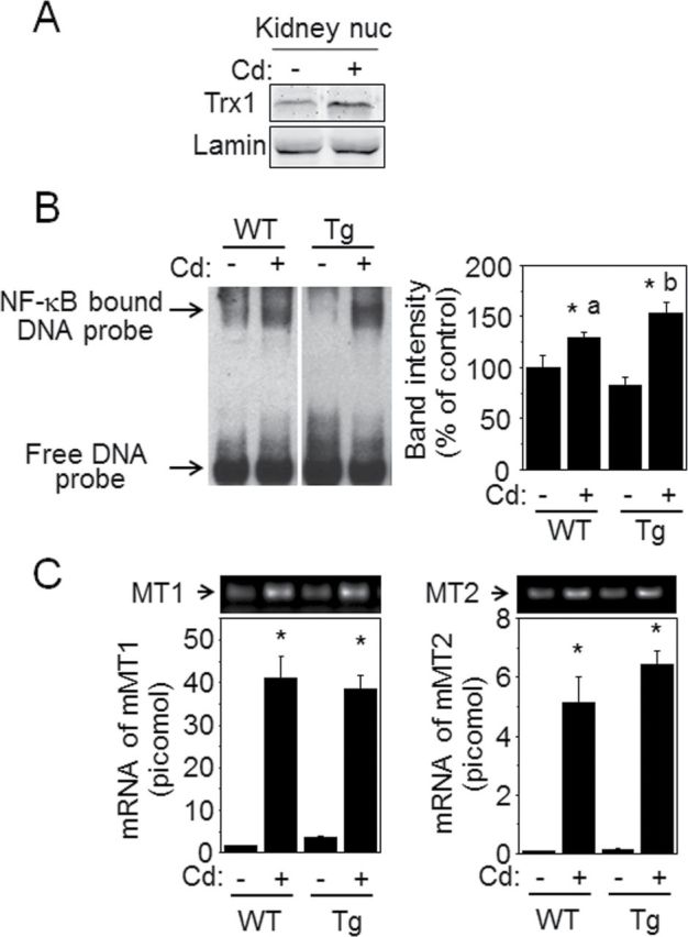
Low level of Cd-induced NF-κB activation in kidney was potentiated by NLS-Trx1 Tg mouse. Kidney tissues obtained from WT and NLS-Trx1 transgenic mice exposed to Cd (10mg/kg) or saline for 6h were used for subcellular fractionation and mRNA isolation. Nuclear fraction was examined for Trx1 nuclear translocation by Western blotting (A) and DNA-binding activity of NF-κB by EMSA (B). (B) Bands indicated as NF-κB-bound DNA probe show activity of NF-κB and intensities of bands were measured as described in Figure 5. Representative results obtained from six mice for each group are shown. *p < 0.05 for WT without Cd. “a” and “b” columns in bar graph (B) show that these are significantly different (p < 0.05). (C) mRNA isolated from kidney tissue was converted to cDNA for qRT-PCR. cDNA was analyzed for MT1 and MT2 by qRT-PCR. Results of mRNA levels for MT1 and MT2 in bar graph are shown as means ± SE. *p < 0.05.
