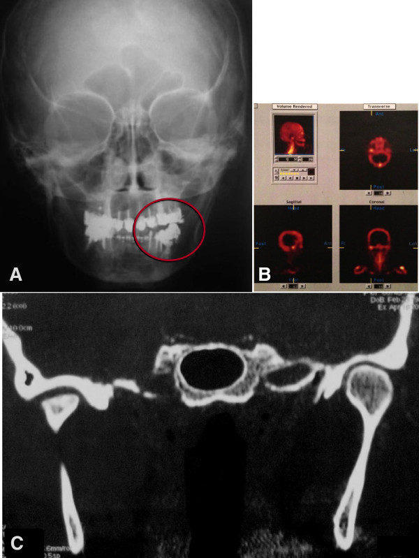
Figure 1.
Preoperative evaluation. (A) Frontal initial pre-operative X-ray (frontal teleradiography): An open bite on the left side was present. (B) Bone scintigraphy: High bone metabolic activity was present at the right TMJ. (C) Computed Tomography in coronal sections of the face: Compared to the left side, the right TMJ presented a substantial loss of condyle anatomy associated with areas of bone sclerosis. The asymmetry between the mandibular ramus marks the lateral deviation of the jaw to the right side.

