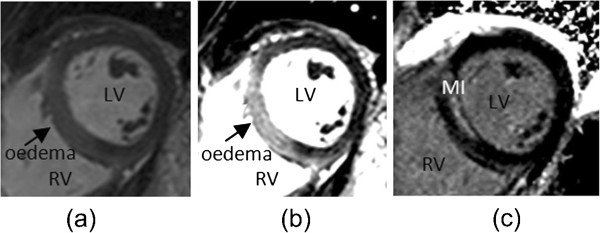
Figure 2.

Example of short axis CMR images: (a) an initial bright blood T2-weighted oedema image and (b) transmural oedema more clearly revealed after user adjustment of window and level settings; (c) the corresponding LGE image reveals non-transmural hyperenhancement in the septum.
