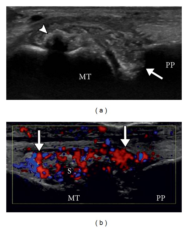
Figure 6.

Gout with synovitis. Longitudinal US images of the 1st MTP joint without (a) and with (b) color Doppler show calcified, shadowing tophus (arrowhead) and adjacent heterogeneous soft tissue with associated hyperemia on color doppler imaging, consistent with synovial proliferation. Note the erosions at base of proximal phalanx (arrow).
