
Figure 2. Modelling the intra-pulse acceleration.

(a) False-colour image (logarithmic scale) of the angularly resolved electron energy distribution at the target rear side, at the time the peak of the laser pulse reaches the front side (t=0). The components of the spatial distribution of the electric field amplitude in target normal direction (Ex), and parallel to the target surfaces (Ey), are plotted in (b) and (c) for t=0, and in (d) and (e) for t=44 fs, respectively. The field distributions are normalized using the maximum field values of 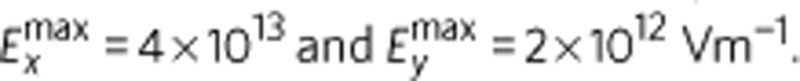 The edge size of the boxes (b–e) corresponds to 18 μm. The trajectories of protons reaching more than 90% of the maximum energy (E>0.9) are overlaid in black. (f) For these protons, the increase of the deflection angle αcent with time is shown in green, together with the evolution of the maximum energy of protons emitted under sample angles α of 6° (blue), −1° (black) and −5° (red). For illustration of the timescale, the laser pulse profile is given by the red shaded region and the black gaussian-shaped curve. For the duration of the laser pulse τ, the asymmetry of the electron distribution translates into a non-target-normal emission of energetic protons that can be detected experimentally.