Figure 4.

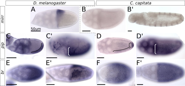
Expression of mirr, pip and br in C. capitata and D. melanogaster oogenesis. All images are in situ hybridizations; posterior is to the right, and ventral to the bottom. The scale bar is always 50 μm. (A)mirr expression in a stage 10 egg chamber of D. melanogaster. (B)mirr expression in a stage 10 egg chamber of C. capitata, with (B’) a positive control for the probe in the embryo. (C) Expression of pip in a stage 9 egg chamber of D. melanogaster shows dorsal and anterior repression of the gene, and an equal expression strength in ventral and posterior follicle cells (marked by bracket). (C’) Stage 10B shows the final stabilized pip pattern. (D) In C. capitata, ventral pip expression starts only at stage 10A, and is visibly lighter than the posterior domain (domains marked by separate brackets). (D’) At stage 10B the pattern has stabilized and shows the same sharp on-off boundary between cells expressing and not-expressing pip as seen in D. melanogaster(C’). (E) Expression of br is visible in all follicle cells of the D. melanogaster stage 9 egg chamber. (E’) Stage 10B shows br expressed in the roof cells of the appendage primordia. (F) In C. capitata, stage 9 expression is similar with all cells expressing br. (F’) A C. capitata stage 10B egg chamber shows how all follicle cells continue expressing br, and a pattern such as in D. melanogaster(E’) is not formed.
