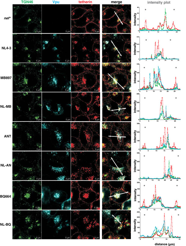
Figure 4.
Cellular localization of tetherin in the presence of various Vpu proteins. Confocal immunofluorescence images of HeLa cells transfected with constructs expressing the indicated Vpu variants. Two days post-transfection, cells were fixed and permeabilized for intracellular staining of tetherin (red), Vpu (blue) and the TGN (green). Images show representative confocal acquisitions of at least 40 transfected cells investigated. Tetherin was located at the cell surface in cells transfected with constructs expressing the parental SIVcpz and SIVgor Vpus but not in cells expressing chimeras containing the TMD of the NL4-3 Vpu. Cellular localization was determined by microscopic examination and by analysis of the Vpu, tetherin and TGN signal intensities throughout the cells (right panel). The regions utilized to generate the profile plots are indicated by the arrows. White circles indicate the position of the plasma membrane.

