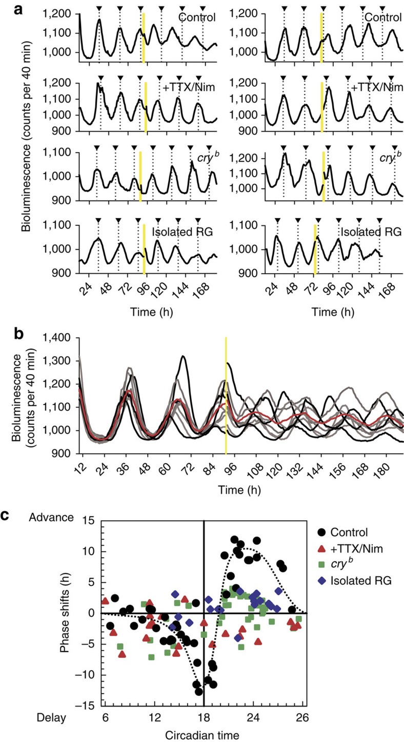
Figure 2. Light-pulse-induced circadian phase-shifts of single PG cells in the CNS–RG complex.
(a) Each trace denotes a representative per-luc rhythm in a single PG cell that received a 30-min light-pulse (yellow bar) immediately after (left) or before (right) the peak of per-luc rhythms. Triangles on the top denote the peak of per-luc rhythms estimated by the waveforms before light-pulse exposure and extended to the end of recordings as a reference. First row: in PG cells in CNS–RG complex cultures, a light-pulse immediately after the peak produced phase-advance and a light-pulse immediately before the peak produced phase-delay in successive per-luc rhythms. Second row: an experiment similar to the above, but CNS–RG complexes were cultured in medium containing TTX (0.3 μM) and nimodipine (Nim; 2 μM). Light-induced phase-advance and -delay were both blocked by TTX and Nim. Third row: an experiment similar to the above, but CNS–RG complexes were cultured from cryb mutant flies. Light-induced phase-shifts were significantly reduced in the cryb mutant. Bottom row: in PG cells in isolated RG cultures from wild-type flies, light-pulse-induced phase-shifts were significantly reduced. (b) Synchronous per-luc rhythms in individual PG cells (grey-scale traces) located in a CNS–RG complex were desynchronized by a 30-min light-pulse given at the approximate middle of the peak of per-luc rhythms. The average amplitude of per-luc rhythms (red trace) was significantly reduced after the light-pulse. (c) Circadian time dependency of light-induced phase-shifts of per-luc rhythms in PG cells. Circadian time 18 was defined by the peak of per-luc rhythms during the pre-exposure period. The second circadian peak after the light pulse was used to estimate phase-shifts. Under control conditions (black circles), light-pulses produced a phase-response curve (hand-fitted curve as dotted line) in which delay shifts switched to advance shifts at circadian time 20. The light-induced phase-responses were reduced by TTX/Nim treatment (red triangles), in PG cells cultured from cryb mutant flies (green squares) and in PG cells from isolated RG cultures (blue diamonds).

