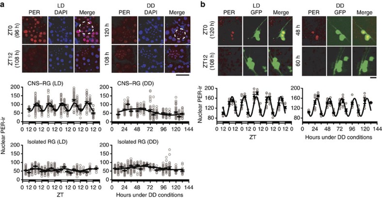
Figure 3. Nuclear PER immunostaining in cultured PG cells.
(a) Images: comparison of anti-PER immunoreactivity (PER-ir) in CNS–RG complexes maintained under 12-h:12-h LD or DD. The time (h) in vitro is indicated on the left. Most PER-ir (red) co-localized with DAPI (blue) nuclear staining at lights-on (ZT0; merged image was purple), but not at lights-off (ZT12), indicated by the diffuse non-nuclear red at ZT12. Cells with small nuclei (<7–8 μm) are CA cells (white dotted line) and cells with larger nuclei (>10 μm) are PG cells. No circadian rhythms in nuclear PER-ir were evident in CNS–RG cultures maintained under DD. Scale bar, 50 μm. Graphs: nuclear PER-ir in PG cells was calculated as the PER-ir/DAPI fluorescence intensity ratio at each time point. Grey circles indicate the level of nuclear PER-ir in each cell (number of cultures, 152; number of cells, 3,173). Nonlinear regression fit a sine wave to the data (solid lines). Robust circadian oscillation of nuclear PER-ir accumulation occurred only in CNS–RG complexes maintained under LD conditions (estimated period, 23.6 h), while non-circadian oscillations occurred in CNS–RG complexes maintained under DD (128.7 h) and in isolated RGs maintained under LD (52.1 h) or DD (122.6 h) conditions. (b) PER-ir in LNs in CNS–RG complex cultures. Images: high levels of PER-ir (red) were observed in LN nuclei at lights-on (ZT0) under LD conditions and during the subjective daytime (48 h under DD conditions). LNs were identified by their expression of GFP in Pdf-gal4/UAS-GFPII S65T flies. Red PER-ir and green GFP image-overlays identified LNs that exhibited rhythmic nuclear PER-ir (yellow in the merge images). Scale bar, 5 μm. Graphs: nuclear PER-ir rhythms under LD cycles exhibited an estimated period of 23.5 h. Free-running nuclear PER-ir rhythms exhibited an estimated period of 24.2 h under DD conditions. In both cases, nuclear PER-ir rhythms were sustained for five circadian cycles in vitro. Nuclear PER-ir in LNs was calculated as the ratio of PER-ir over averaged background fluorescence intensity.

