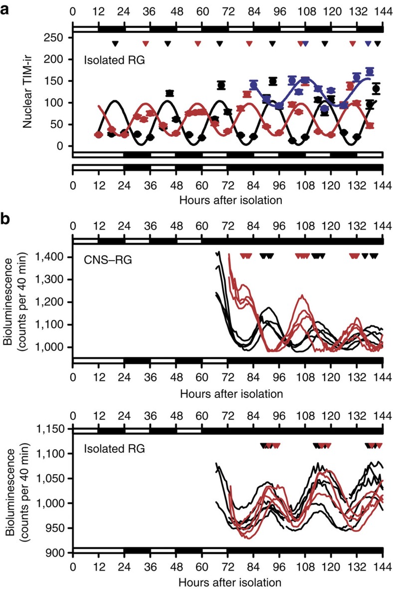
Figure 5. Selective entrainment of nuclear TIM expression rhythms to LD cycles in PG cells in isolated RGs.
(a) Nuclear TIM-ir rhythms in PG cells in isolated RG cultures maintained under standard LD cycles (black circles), reversed LD cycles (red circles), and DD following the reversed LD cycles (blue circles). Peak TIM-ir rhythms are marked on the top by triangles in corresponding colours. TIM-ir rhythms in PG cells were synchronized to the LD cycles in the isolated RG and had free-run under DD with an initial circadian phase coupled to the precedent LD cycles. (b) Representative per-luc rhythms in PG cells, which was cultured under standard LD cycle conditions (black traces) or under reversed LD cycle conditions (red traces) before recordings under DD conditions. Peak of per-luc rhythms estimated in PG cells pre-cultured under standard LD cycle conditions (black triangles) and under reversed LD cycle conditions (red triangles) were marked on the top. Per-luc rhythms in PG cells synchronized to the LD cycles in the CNS–RG complex but not in the isolated RG.

