Fig. 5.
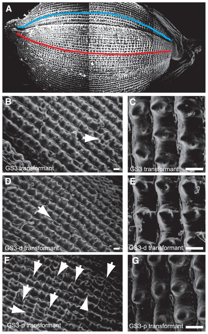
Scanning electron micrographs of the upper epidermis in the transgenic plants. (A) The three sequential images of a seed were merged into a single image. The cell numbers of the upper epidermis were counted in the longitudinal tubercles that are formed from the apex to the base of a seed. Red and blue lines indicate a row of longitudinal tubercles at the lemma and palea, respectively. (B, C) The upper epidermis in the GS3 transformant. (D, E) The upper epidermis in the GS3-d transformant. (F, G) The upper epidermis in the GS3-p transformant. Arrows indicate the irregular thin rows. Bars are 50 μm.

