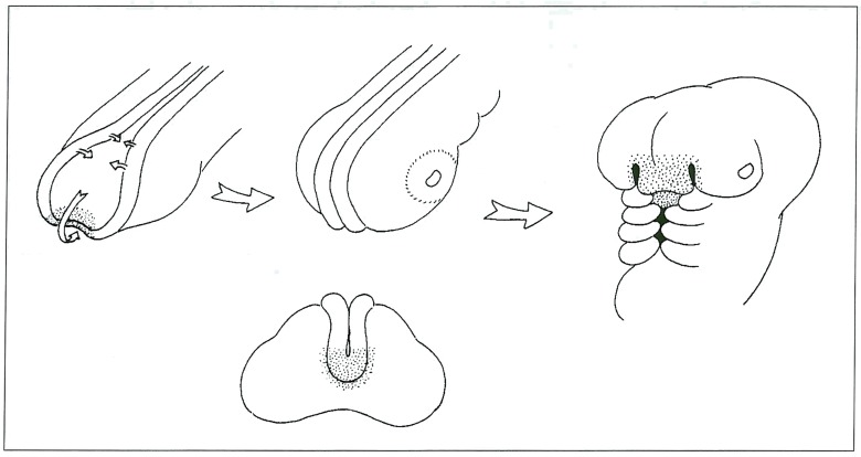
Figure 5.
A) Drawing of the rostrum of an embryo, the arrows indicate progressive closure of the neural folds and curling inferiorly to the anterior lip of the neuropore. Stippled area indicates anlage of the hypothalamus, adenohypophysis and covering nasal tissues. B, C) Oblique and frontal views showing migration of this region. D) Later embryo showing nasal tissues in their final location. Future hypothalamus and adenohypophysis are still just visible prior to closure of the maxillary buds.

