Fig. 7.
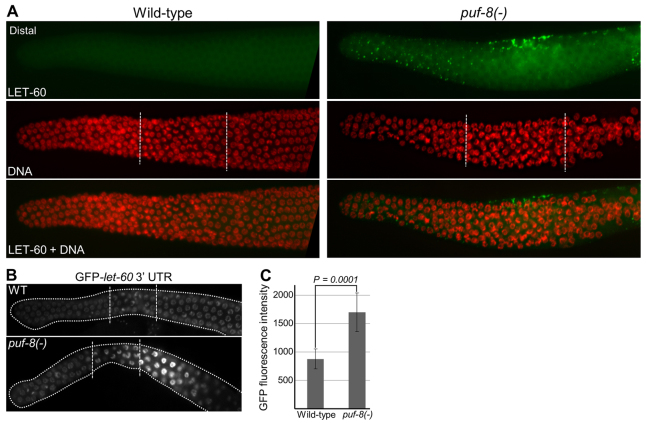
PUF-8 negatively regulates LET-60 expression in the germline. (A) Dissected gonads stained with anti-LET-60 antibodies and DAPI. Genotypes are indicated on the top. In the transition zone, although LET-60 signal is seen only in a few cells at the top in this focal plane, it was observed in a several cells within this zone in other focal planes. The dots revealed by immunostaining signal correspond to cell membrane at the intersection of adjacent cells. See also supplementary material Figs S5 and S6. (B) Dissected gonads of live worms revealing the expression pattern of GFP::H2B reporter fusion expressed under the control of pie-1 promoter and let-60 3′ UTR sequences. Only the distal germlines are shown. In the puf-8(-) genetic background, let-60 3′ UTR fusion shows significant upregulation in the transition and early pachytene region when compared with the wild type. In total, 20 worms each of 11 independent transgenic lines were examined, and representative images are shown here. broken lines in A and B mark the boundaries among the mitotic, transition and pachytene regions. (C) Comparison of the GFP fluorescence intensity (arbitrary units) per nucleus in the transition zone between the wild-type and puf-8(-) gonads. n=23 nuclei from five gonads; P=0.0001 (Student’s t-test).

