
Fig. 3.
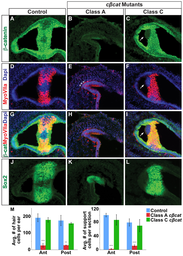
Vestibular hair and support cells are dependent on canonical Wnt signaling. (A-C) Immunostaining for β-catenin on sections through the crista ampullaris (anterior or posterior) in control (A) and cβcat mutant (B,C) mice at E14.5 (tamoxifen administered at E10.5). β-catenin expression is greatly reduced in Class A mutants and only occasionally affected in Class C mutants, as compared with controls. (D-L) The numbers of myosin VIIa+ hair cells and Sox2+ support cells (J-L) are greatly reduced in Class A mutants but only occasionally affected in Class C mutants. Asterisks (E) mark examples of the few myosin VIIa+ cells detected in Class A mutants. Arrows (C,F,I) highlight the cell-autonomous loss of myosin VIIa staining in β-catenin-deficient cells in Class C mutants. (M) Quantification of hair and support cells in control (blue), Class A (red) and Class C (green) mutants. Error bars indicate s.e.m. ***P<0.01, **P<0.05 (unpaired Student’s t-test).