Fig. 4.

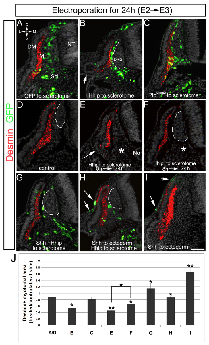
Misexpression of Hhip in avian sclerotome represses the myogenic activity of No/floor plate-derived Shh. Transverse sections of embryos for which ventral somites were electroporated at E2 and fixed 24 hours later. (A-F) Control GFP (A), Hhip/GFP (B) or PTCΔloop2 (C) embryos. Note in B absence of a lateral myotome with concomitant looping of the VLL (arrow). (D-F) Co-electroporation to the ventral somite (asterisk) of TRE-Hhip+rtTA2s-M2 followed either by immediate activation of expression with Dox (E) or delayed activation after 8 hours (F). Note the small and intermediate sizes of the myotomes in E and F, respectively, compared with controls (A,D). The lateral myotome is virtually absent in E, as in B (arrows). (G-I) The effect of Hhip is rescued by excess Shh. (G) Embryos co-transfected with Shh and Hhip to sclerotome. (H) Sequential electroporation of Shh to ectoderm (arrows) followed by Hhip to sclerotome. (I) Electroporation of Shh alone to ectoderm (arrows). In addition to myotomal expression, desmin is sometimes apparent in the basal side of the medial DM (D), where it is particularly upregulated following local Shh application (H,I). (J) Quantification of the area occupied by desmin+ myotomes in treated compared with intact contralateral sides. Letters under bars refer to treatments/groups described in A-I. Error bars represent s.e.m. *P<0.02, **P<0.0001. Dorsal root ganglia (DRG) are marked by a dashed line. Scale bar: 100 μm. DM, dermomyotome; M, myotome; NT, neural tube; Scl, sclerotome.
