Fig. 2.
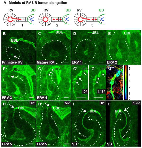
Three-dimensional nephron lumen elongation. (A) Models of nephron lumen elongation. The renal vesicle (RV) lumen may extend towards the ureteric bud (UB) lumen (1), additional de novo lumens may form and coalesce (2), or a combination of extension and de novo lumen generation may exist (3). (B-I′) Three-dimensional reconstructions from confocal z-stacks of immunostaining with aPKC (green) and NCAM (not shown). aPKC marks the apical surface, thereby demarcating the lumen. (B,C) A primitive renal vesicle (B) (PRV) has one or two small lumens that expand to form a mature RV with a single expanded lumen (C). (D) The lumen extends distally towards the ureteric bud lumen (UBL) in stage 1 of the extended renal vesicle (ERV1). (E,F) Lumen extension progresses (ERV2) (E), and then de novo lumen formation (arrow) occurs in the distal or connecting segment of ERV3 (F). (G) Additional distinct lumens (arrows) form in the connecting segment (ERV4). (G′,G″) High magnification of the connecting region shows separation between lumens (arrows) at 0° (G′) and 148° (G″) rotation. (G″′) A depth plot of 0-5 μm illustrates that one of the lumens (asterisks) is not at the same depth as neighboring lumens, which emphasizes its unique origin. (H,H′) Connecting segment lumens begin to coalesce in ERV5, yet the ERV5 lumen is not fused to the UBL. The ERV5 lumen appears fused to the UBL when viewed at 0° rotation (H), but is clearly separate when the 3D reconstruction is rotated to 56° (arrow, H′). (I,I′) As the proximal lumen curves to form an S-shaped body (SB) there is one continuous lumen (arrows). A 3D SB at 0° (I) and 136° (I′) rotation is shown. Results are representative of sections from six mice. Scale bars: 10 μm in B-I′.

