Fig. 3.
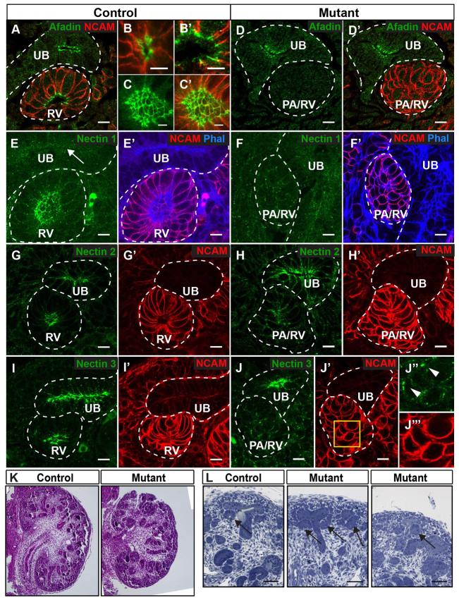
Afadin is required for nectin clustering in renal vesicles. (A-J) Localization of afadin (A-D′), nectin 1 (E-F′), nectin 2 (G-H′) and nectin 3 (I-J″′) in E14.5 kidneys from control and mutant are indicated (green). NCAM (red) delineates nephron precursors and their derivatives; phalloidin (blue) stains F-actin. (A) Afadin is expressed in ureteric bud (UB) and renal vesicles (RV). (B,B′) High magnification of a primitive (B) and mature (B′) RV is shown. (C,C′) Imaging of the lumen en face in mature RVs shows afadin at apical lateral cell junctions and NCAM at the basolateral surface in mature RVs. (D,D′) Mutant kidneys lack afadin in PA/RV. (E-F′) Nectin 1 is expressed in RV. In mutants (F,F′), nectin 1 is not recruited to an apical surface in pretubular aggregates/renal vesicles (PA/RV). (G-J) Nectin 2 and 3 are expressed at the apical lateral surface of UB and RV. In mutants, nectin 2 and 3 are not recruited to an apical surface in PA/RV. (J′-J″′) The inset in J′ (J″,J″′) shows nectin 3 (green) at discrete cell surface patches in mutant PA/RV. All mutant panels show absence of a central clearing or lumen in PA/RVs. (K,L) Hematoxylin and Eosin-stained (K) and Toluidine Blue-stained (L) sections from control (Afadinfl/fl) and mutant (Afadinfl/fl; Pax3-cre) kidneys at E14.5. Arrows in L indicate PA/RV. Lumens are absent in mutants. Broken lines indicate UB and PA/RV. All results are representative of sections from three mice. Scale bars: 5 μm in B-C′; 10 μm in A,D-J′; 50 μm in L.

