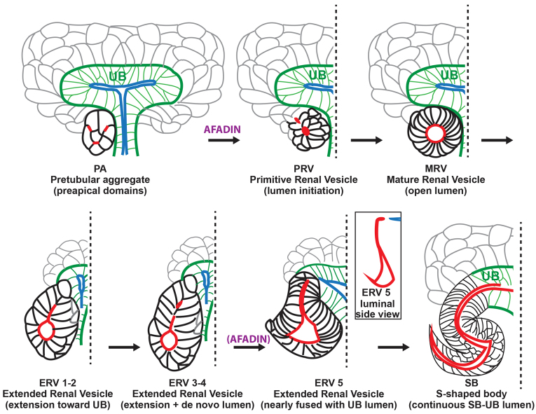
Fig. 7.
New model of nephron lumen formation. In pretubular aggregates (PA), Par3-containing pre-apical domains (red) are present in numerous membranes. These domains reorganize and coalesce in an afadin-dependent manner to form one or two small apical foci/lumen in primitive renal vesicles (PRV). Par3 is redistributed to apical lateral cell junctions (not illustrated) and the lumen expands in the mature renal vesicle (MRV). Next, the expanded lumen (red) extends towards the ureteric bud lumen (blue) in the extended renal vesicle (ERV) and then additional de novo lumens form in the distal segment. In the late ERV, these additional distal lumens coalesce, probably in an afadin-dependent manner, whereas the proximal region of the lumen extends. Finally, the extended lumen joins the ureteric bud lumen at the S-shaped body stage (SB), where a pronounced S-shaped lumen is visible.

