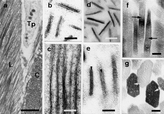
Fig. 1.

Electron micrographs of the processes of crystal development in normal enamel. (a) Ribbon-shaped structures and the Tomes’ processes (Tp) of ameloblasts at the secretory stage. Cross-(C) and longitudinal sections (L) of early ribbon-shaped structures. (b) Higher magnification of cross-sections of early ribbon-shaped structures (double-stained sections). (c) Longitudinal sections of early ribbon-shaped structures. A ribbon-shaped structure consists of 2 thin organic layers and an electron-lucent mineral zone (double-stained sections). (d) Higher magnification of cross-sections of early ribbon-shaped structures. Precursor minerals without lattice lines are observed as electron-dense fuzzy images (unstained sections). (e) The appearance of the initial lattice line (arrowheads) within the envelope (double-stained sections). (f) Developing longitudinal sections of enamel crystals with central dark lines (arrows, unstained sections). (g) Developing cross-sections of enamel crystals with central dark lines (arrows, unstained sections). Scale bars: (a) 1 μm, (b–d) 20 nm, (e–g)) 10 nm.
