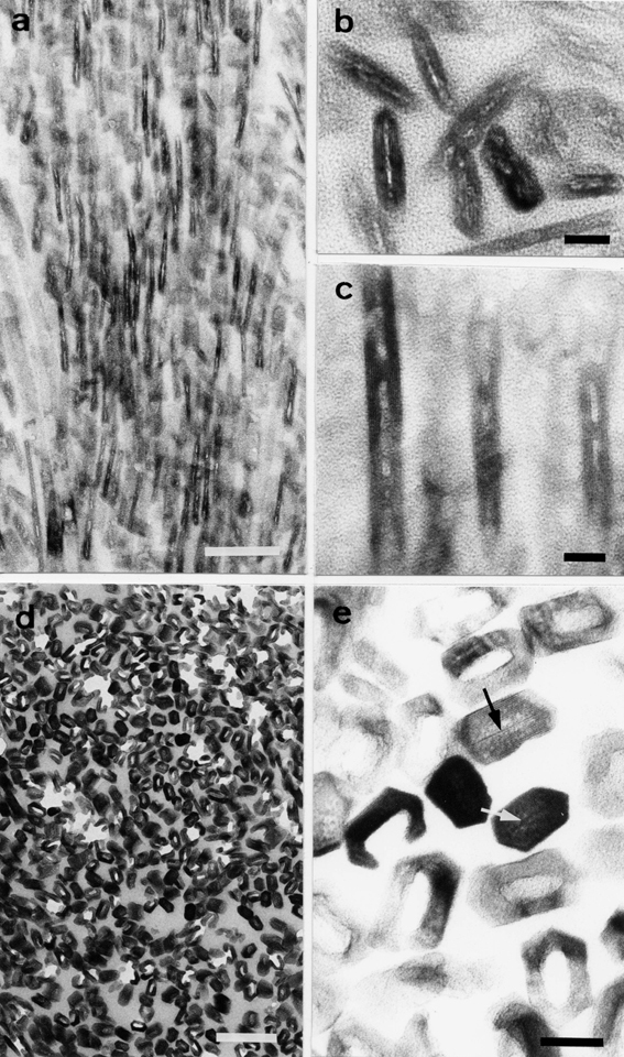
Fig. 2.

Electron micrographs of crystals obtained from cadmium-exposed rat tooth enamel. (a) Low magnification of enamel crystals affected by the cadmium exposure for 5 weeks. (b and c) Higher magnification of enamel crystals. The crystal nucleation process seems to be sporadically inhibited. (d and e) Enamel crystals affected by the cadmium exposure for 12 weeks. (d) Perforated crystals at low magnification. (e) Cross-section of perforated and intact crystals at higher magnification. The perforated crystals reveal voids at their centers, while the intact crystals show central dark lines (arrowheads). Scale bars: (a and d) 100 nm, (b, c and e) 10 nm. (a–e) Unstained sections.
