
Figure 1.
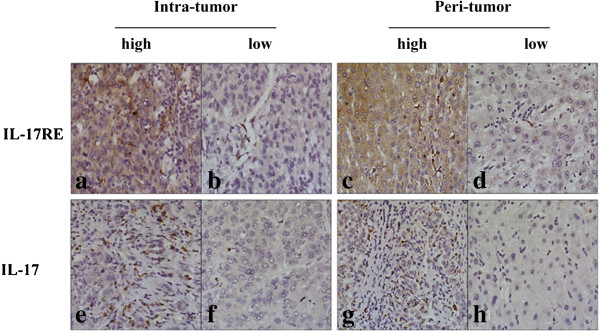
Immunohistochemistry analysis of IL-17RE and IL-17. a-h showed high (a, c, e and g) and low (b, d, f and h) densities of IL-17RE and IL-17 staining cells in intratumoral (a, b, e and f) and peritumoral area (c, d, g and h), respectively (x 200).