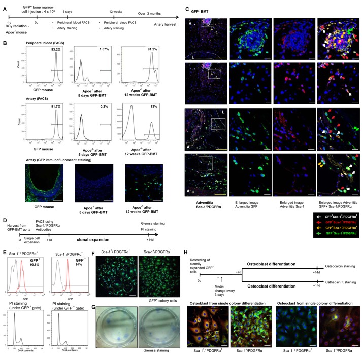
Figure 3. The origin of calcifying progenitor cells.
(A) BMT experimental outline. (B) After 5 d of cell infusion, we checked the presence of GFP+ cells in arteries and blood using FACS and immunofluorescent staining. A small fraction of donor GFP+ cells were detected in peripheral blood (1.5%) but were rarely detected in the artery (0.2%). Twelve weeks after transplantation GFP+ cells from donor BM were reconstituted with peripheral blood cells of C57 mice (up to 90%). At that time, 13% of arterial resident cells were GFP+. Thus, the majority of GFP+ cells gradually were incorporated into the artery within a considerable duration. Bars: 50 µm. (C) Aortas were harvested after 6 mo of BMT and were stained with antibodies targeting GFP, Sca-1, or PDGFRα. The three panels on the right depict high-magnification images of the white squares shown on the left. Blue, Sytox Blue nuclear staining; L, lumen; A, adventitia. White dashed lines describe the media. Bars: yellow = 100 µm; white = 20 µm. (D) Schematic of the GFP+ clonal expansion assay. (E) PI staining to identify fusion between GFP+ and non-GFP+ cells. (F) GFP+ single clone immunofluroscent staining. Bars: 200 µm. (G) Giemsa staining of single-cell colonies. (H) Osteocalcin/cathepsin K staining of osteoblast/osteoclast differentiation from GFP+ single colonies after 14 d.

