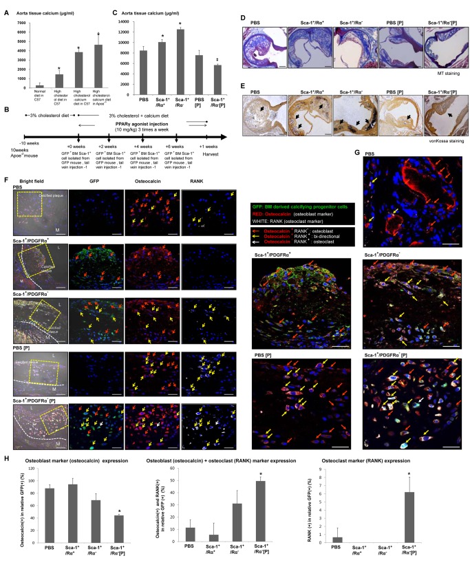
Figure 7. Calcifying progenitor cells induce calcification in atherosclerotic plaques of Apoe−/− mice.
(A) Calcium accumulation levels in mouse arteries according to diet. *P<0.005 versus C57 fed a normal diet (n = 10 per group). (B) Experimental timeline. (C) Calcium accumulation levels in the aortas of Apoe−/− mice according to BM cell type injected and PPARγ activation. *P<0.001 versus PBS-treated group. ‡P<0.001 versus mice injected with BM Sca-1+/PDGFRα− cells. (D) Atherosclerotic plaque with MT staining. Bars: 200 µm. (E) Calcification induction in atherosclerotic plaques was detected by von Kossa staining. Arrows indicate calcified areas. Bars: 200 µm. (F) Atherosclerotic calcification plaque immunostained with osteocalcin, an osteoblastic marker, and RANK, an osteoclastic marker, to determine the fates of infiltrating BM-derived calcifying progenitor cells in the presence/absence of PPARγ activation. Bars: 20 µm. (G) Image enlargement showing osteocalcin and RANK immunostaining. Bars: 20 µm. (H) Osteoblastic, osteoclastic, and bidirectional cell counts in the artery. GFP+Sca-1+/PDGFRα+ cells primarily expressed osteoblast markers. GFP+Sca-1+/PDGFRα− cells mainly differentiated into osteoblast-like cells, but some infiltrating cells differentiated into bidirectional cells. When PPARγ was activated, differentiating GFP+Sca-1+/PDGFRα− cells shifted from osteoblast-like to bidirectional cells or osteoclasts (n = 10 per group). *P<0.005 compared to mice injected with BM-derived Sca-1+/PDGFRα− cells. P, PPARγ agonist.

