
Figure 3.
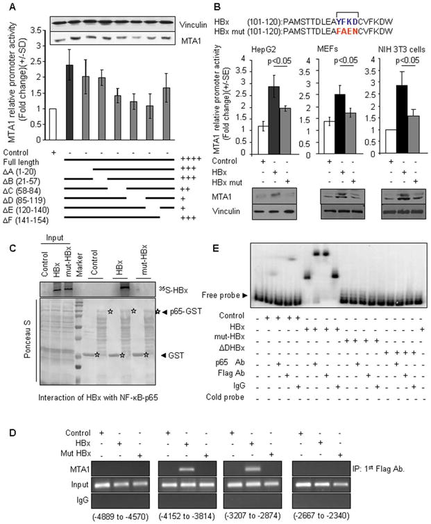
Role of YFKD motif of HBX in regulation of MTA1 transcription. (A) MTA1 promoter activity in HepG2 cells transfected with either control vector or HBx expression vector or HBx-deletion constructs. Inset: Western blot analysis of MTA1 protein in HepG2 cells after transfecting with vector or HBx or HBx- deletion constructs. (B) MTA1 promoter activity in HepG2 or NIH 3T3 or MEF cells after being transfected with either control vector or HBx expression vector or mut-HBx (YFKD was mutated to FAEN) expression vector. Lower panels are Western blot analysis of MTA1 protein in HepG2 or NIH 3T3 or MEF cells after being transfected with either control vector or HBx expression vector or mut-HBx expression vector. (C) GST pulldown assays with 35S-labeled in vitro-translated HBx and mut-HBx and GST- NF-κB-p65 protein. (D) Recruitment of HBx to MTA1-chromatin (−3814 to −4152 and −2874 to −3207) by ChIP assay in the NIH 3T3 cells. (E) EMSA analysis of NF-κB-p65 binding to the mouse MTA1 promoter using PCR product encompassing functional NF-κB consensus sequence in transfected cells and controls. Nuclear extract of HepG2 cell transfected with either Flag-tagged HBx, or Flag-tagged mut-HBx, or Flag-tagged ΔD-deletion HBx (2000 ng/lane), probe control (0.3 ng/lane), NF-κB-p65 antibody (Ab.), anti-Flag antibody, and IgG control (1000 ng/lane), cold probe (15 ng/lane) were used.