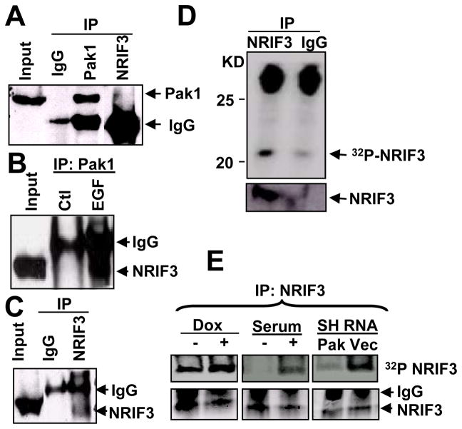
Figure 2. NRIF3 is a Pak1-interacting substrate in a Pak1-sensitive manner.
(a–c) NRIF3 binds with Pak1 in vivo. Total lysates were made from MCF-7 cells treated with or without EGF overnight, immunoprecipitated with IgG, Pak1, or NRIF3 antibodies, and resolved on SDS-PAGE. Western blot analysis was performed using antibody against Pak1 or NRIF3. (a) Pak1 binds with NRIF3 (4th lane from left; 1st and 3rd lane as positive control). (b) NRIF3 binds strongly with Pak1 in presence of EGF (3rd lane, lower band). (c) NRIF3 can be detected by self-immunoprecipitation. (d) NRIF3 is phosphorylated in vivo. MCF-7 cells were labeled with 32P orthophosphate and immunoprecipitated with NRIF3 antibody or IgG, resolved on SDS-PAGE, transferred to a blot, and examined by autoradiography or Western blot analysis using anti-NRIF3 antibody. Upper panel is the autoradiogram and lower panel is the Western blot analysis. (e) Over-expressing Pak1 clones (left panel), MCF-7 cells with or without serum (middle panel), Pak1 silencing SH-RNA Pak1 or vector clones were labeled with 32P orthophosphate and immunoprecipitated with NRIF3 antibody. Upper panels show the change in NRIF3 phosphorylation as Pak1 is included or depleted.

