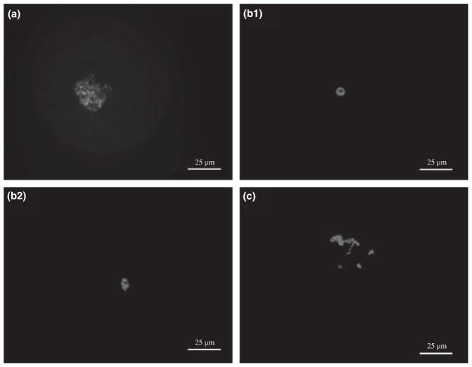
Fig. 1.
Identification of chromatin configurations in GV-stage ferret oocytes. (a) Fibrillar chromatin (FC) – chromatin fibrils are distributed throughout the GV. (b1 and b2) Condensed chromatin (CC) – (b1) circular or (b2) oval form of highly condensed chromatin surrounds the nucleolus. (c) Intermediate condensed chromatin (ICC) – dense chromatin masses within the GV. Ocytes were stained with DAPI and examined by fluorescence microscopy. Bar = 25 μm

