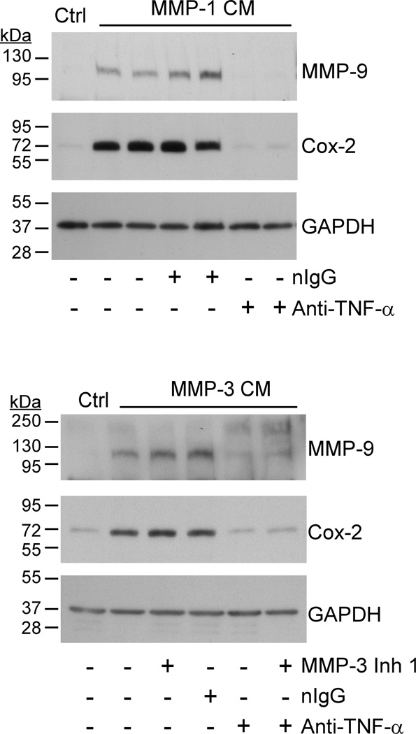
Figure 7. Neutralizing anti-TNF-α IgG blocks MMP-induced Cox-2 expression.
Conditioned media recovered from RAW264.7 macrophages (24–well plate; 2.5 × 10⁵/well) exposed (1 h) to 50 nM MMP-1 were subsequently incubated 2 h with non-immune IgG1 (20 µg/ml) or rat monoclonal anti-mouse TNF-α IgG1 (20 µg/ml). Conditioned media and antibody treated conditioned media were then added to naïve macrophages in duplicate wells. In a similar experiment, conditioned media recovered from cells incubated with MMP-3 were treated with MMP-3 Inhibitor 1 (20 µM) and/or anti-TNFα (20 µg/ml) prior to adding to naïve macrophages. Macrophages were incubated with conditioned media overnight and lysates prepared. Levels of Cox-2 and GAPDH in lysates were determined by Western blot. The data are representative of three separate experiments.

