Fig.1.
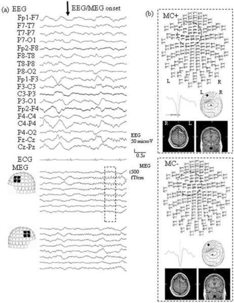
(a) Electroencephalography (EEG) and magnetoencephalography (MEG) waveforms at electrical/magnetic seizure onset, i.e. before any head movement.
(b) Result of dipole analysis with/without movement compensation (MC) of 4 spikes during the time enclosed by the dotted line in Fig. 1a. Top view images of spikes (upper panel), representative spike waveforms (middle left), MEG contour map (middle right), and axial and coronal MRIs of the patient's with spike dipoles (lower) are shown. The dotted crcles on the top view images highlight the sensors showing maximal epileptic activity.
Note that, both with and without MC, the spike distribution as well as MEG spike dipole locations are concordant on a sublobar level to the left mid frontal gyrus.

