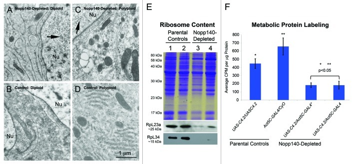
Figure 2. TEM, immuno-blot analysis and metabolic protein labeling demonstrate loss of ribosomes and protein synthesis. (A−D) Separate tissues were isolated by hand and prepared for TEM analysis. Imaginal wing disc cells from Act5C > UAS-C4.2 larvae (A) contained fewer ribosomes as compared with similar cells from wild type (w1118) larvae (B). Polyploid midgut cells from Act5C > UAS-C4.2 larvae (C) contained far fewer ribosomes than did similar, identical cells from wild type (w1118) larvae (D). Nuclei (Nu) in Act5C > UAS-C4.2 larvae (panels A and C) contained numerous copia virus-like particles indicating stress. Scale bar: 1 µm for all TEM images. (E) Specific antibodies showed a loss of RpL23a and RpL34 in lysates from Act5C > UAS-C4.2 larvae compared with parental type larvae. Lane 1: homozygous UAS-C4.2 parental lysate; Lane 2: heterozygous Act5C-GAL4/CyO-GFP parental lysate; Lane 3: RNAi-expressing UAS-C4.2/Act5C-GAL4 lysate; Lane 4: a separate RNAi-expressing UAS-C4.2/Act5C-GAL4 lysate. (F) Metabolic protein labeling was significantly reduced in Nopp140-RNAi-expressing UAS-C4.2/Act5C-GAL4 larvae compared with larval parental controls. The entire experiment was repeated three times. Absorbance values were determined in triplicate. Graphs were assembled using Microsoft Excel; two-tailed P values were equated using t-Test analysis (two sample assuming unequal variances).

An official website of the United States government
Here's how you know
Official websites use .gov
A
.gov website belongs to an official
government organization in the United States.
Secure .gov websites use HTTPS
A lock (
) or https:// means you've safely
connected to the .gov website. Share sensitive
information only on official, secure websites.
