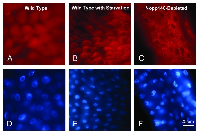
Figure 5. Autophagy was prominent in larval polyploid cells depleted for Nopp140. (A and D)da > mCherry-ATG8 in a wild type background. Labeling remained diffuse throughout the cell. (B and E) As a positive control for autophagy, similar da > mCherry-ATG8a larvae were starved of amino acids. The accumulation of mCherry-ATG8a into autophagosomes and lysosomes (red speckling in the cytoplasm) indicated autophagy as expected due to starvation. (C and F) Well fed da > UAS-C4.2; mCherry-ATG8a larvae displayed similar accumulations of mCherry-ATG8a into cytoplasmic vesicles within their midgut cells indicating premature autophagy in response to the loss of Nopp140.

An official website of the United States government
Here's how you know
Official websites use .gov
A
.gov website belongs to an official
government organization in the United States.
Secure .gov websites use HTTPS
A lock (
) or https:// means you've safely
connected to the .gov website. Share sensitive
information only on official, secure websites.
