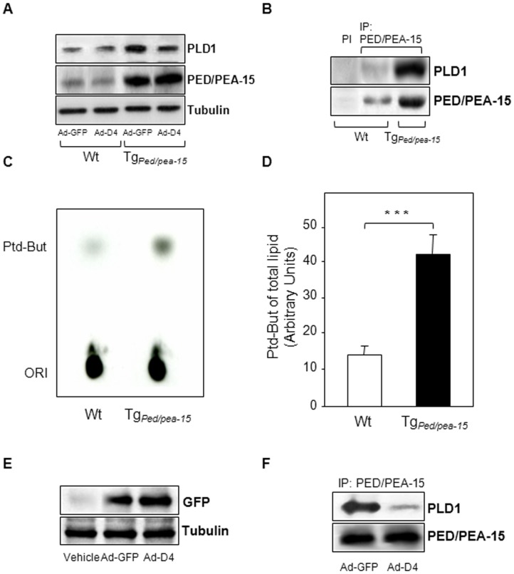
Figure 3. Protein interaction of PED/PEA-15 with PLD1 and PLD1 activity in TgPed/pea-15 mice.
A) Immunoblots of total protein lysates from skeletal muscle homogenates of wild type (Wt) and TgPed/pea-15 mice at one week post Ad-D4 or Ad-GFP infection. The blots were probed with anti-PLD1, anti-PED/PEA-15 and anti-Tubulin antibodies. PI stands for pre-immune serum. B) Immunoblots of immunoprecipitated from skeletal muscle homogenates of wild type (Wt) and TgPed/pea-15 mice. IP were performed using the anti-PED/PEA-15 antibody as described in Experimental Procedures. The upper blot was probed with anti-PLD1 antibody, while the bottom blot was striped and then probed with anti-PED/PEA-15 antibody. C) PLD1 activity was analyzed in skeletal muscle homogenates from wild type (Wt) and TgPed/pea-15 mice by measuring the transphosphatidyl-butanol levels as described in Materials and methods. The autoradiography shown is representative of three independent assays. D) The bar graph represents the densitometric quantization of the spots in three experiments in triplicate. ***p<0.001 vs. Wt. E) Immunoblots of whole lysates from skeletal muscle homogenates of TgPed/pea-15 mice at one week post Ad-D4 or Ad-GFP infection or PBS injection (vehicle). The upper blot was probed with anti-GFP antibody, while the bottom blot was striped and then probed with anti-tubulin antibody. F) Immunoblots of immunoprecipitated from skeletal muscle homogenates of TgPed/pea-15 mice at one week post Ad-D4 or Ad-GFP infection. IP were performed using the anti-PED/PEA-15 antibody. The upper blot was probed with anti-PLD1 antibody, while the bottom blot was striped and then probed with anti-PED/PEA-15 antibody.

