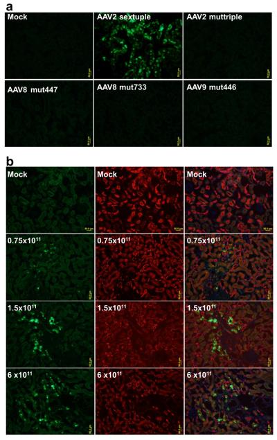
Figure 1.
GFP Expression in the kidneys. a, Strong green fluorescence was observed in renal tubular cells with rAAV2-sextuple, while faint background auto-fluorescence was seen in the PBS and other AAV mutants injected kidney. b, Kidney sections from dose response experiment were contained with GFP (green) and tubular markers(PNA for distal and collecting duct, red). Scale bar: 50 μm.

