
Figure 3. Overnight food deprivation alters the profile of synaptic inputs to DMH cholinergic neurons.
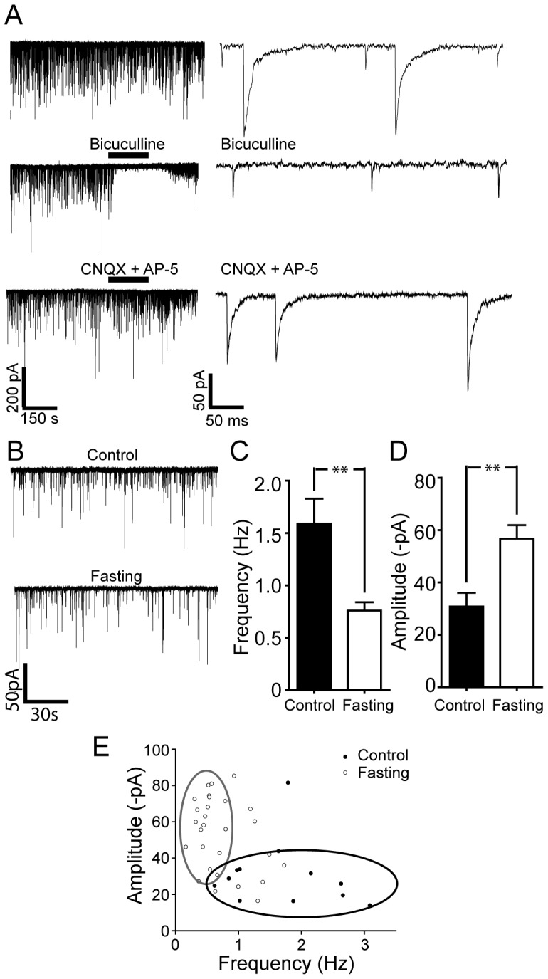
A. Representative recordings of sPSCs activity in identified cholinergic neurons. Two components of PSCs were evident at a holding potential (HP) of −70 mV (upper right panel: on the expanded time scale). Middle panel shows that the GABAA receptor antagonist, bicuculline completely blocked slow synaptic currents (middle panel; right: expanded time scale), while the glutamate receptor antagonists abolished fast synaptic currents (bottom panel, right: expanded time scale). Thus, the DMH cholinergic neurons received both GABAergic and glutamatergic currents. B. Synaptic activity in DMH cholinergic neurons under control and post-fasting conditions. Sample traces show typical examples of the spontaneous synaptic currents recorded in cholinergic neurons under control vs. overnight fasting conditions. C and D. Pooled data of the frequency (C) versus the amplitude (D) of sPSCs from 13 and 29 DMH cholinergic neurons in control and fasting groups. Overnight fasting significantly modulated the frequency as well as the amplitude of sPSCs. E. Plot of amplitude vs. frequency values for sPSCs under control vs. overnight fasting conditions.