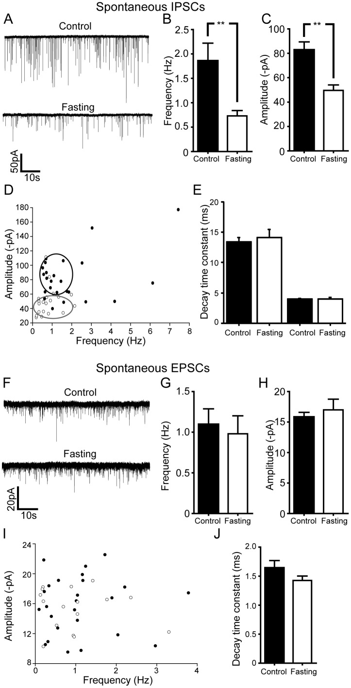
Figure 4. Overnight food deprivation results in significant reductions in net inhibitory tone to DMH cholinergic neurons.
A. Spontaneous GABAergic (IPSC) activity in DMH cholinergic neurons under control vs overnight fasting conditions. There was a significant decrease in the GABAergic transmission following overnight fasting. B and C. Summary plots of the changes in frequency (control: n = 24 neurons, Fasting: n = 19 neurons) and amplitude (control: n = 24 neurons, Fasting: n = 19 neurons) of GABAergic sIPSCs in DMH cholinergic neurons following overnight fasting conditions. Both parameters were significantly decreased. D. Pooled data showing the frequency versus the amplitude of sIPSCs. Food deprivation decreased both the frequency and the amplitude of sIPSCs recorded in DMH cholinergic neurons (Filled circle, control; open circle, fasting). E. Graph showing no significant difference in the decay time constant of sIPSCs in the DMH cholinergic neurons under control and fasting conditions. F. Sample recording traces of pharmacologically isolated glutamatergic s EPSCs in control vs. post-fasting conditions. G and H. Summary plots showing the frequency and the amplitude of sEPSCs in control and fasting groups. There was no change in both parameters. I. Pooled data comparing the frequency vs. the amplitude of sEPSCs in DMH cholinergic neurons under control (filled circle) vs. post fasting (open circle) conditions. J. Graph shows no difference in the decay time constant of sEPSCs in control and fasting groups.

