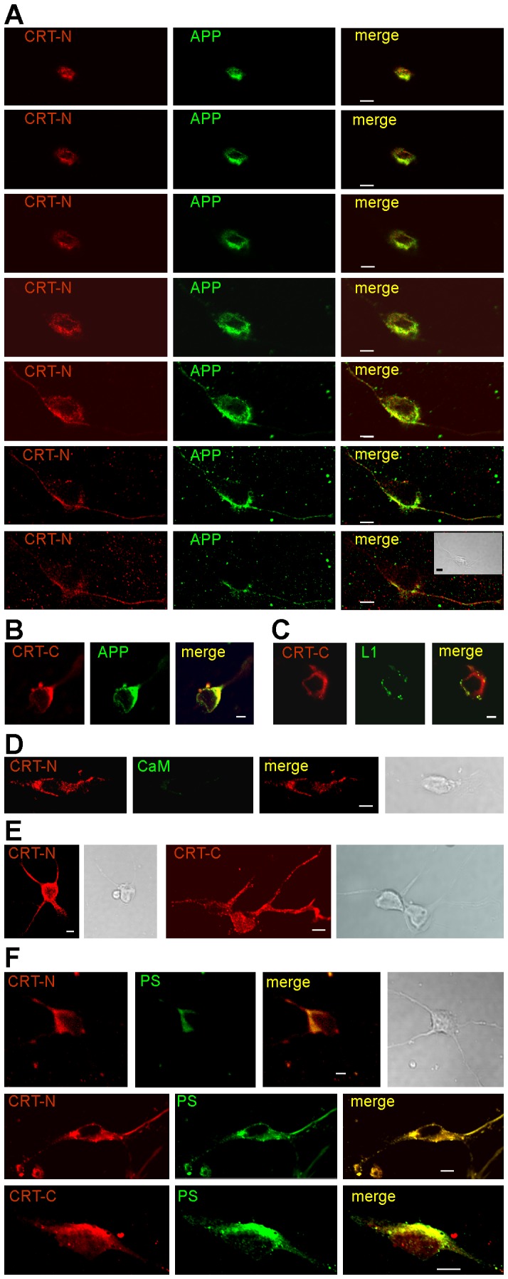
Figure 6. Co-staining of calreticulin with APP and presenilin at the cell surface of live hippocampal neurons.

Live hippocampal neurons were incubated with goat calreticulin antibody T-19 recognizing the N-terminus (CRT-N) (A, D, F) or C-17 recognizing the C-terminus (CRT-C) (B, C, F) and with rabbit APP antibody A8967 directed against the N-terminus (A, B), rabbit L1 (C) or calmodulin (CaM) (D) antibody. After fixation, cells were incubated with the calreticulin antibodies (E) or the rabbit presenilin antibody 2953 and fluorescent-labeled secondary antibodies. Superimposition of immunostainings (merge) shows co-localization of calreticulin and APP (yellow). Phase contrast shows the cellular structures. Scale bar, 5 µm.
