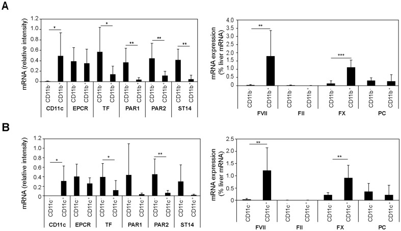
Figure 6. Expression analysis of PyMT tumor macrophages.
PyMT tumors from heterozygous PyMT-EPCRLow/WT mice were dispersed and macrophages were separated with αCD11b (A) or αCD11c (B) paramagnetic beads from tumor cells and stromal cells. Tumor macrophages were CD11b+/CD11c+/F4/80+ by FACS and CD11c was used to assess the efficiency of selection. Expression of the indicated mRNAs was determined by RT-PCR. Coagulation factor mRNA was normalized to a standard curve of normal mouse liver mRNA (* p<0.05, ** p<0.005 T-test, mean±SD, n = 5–8).

