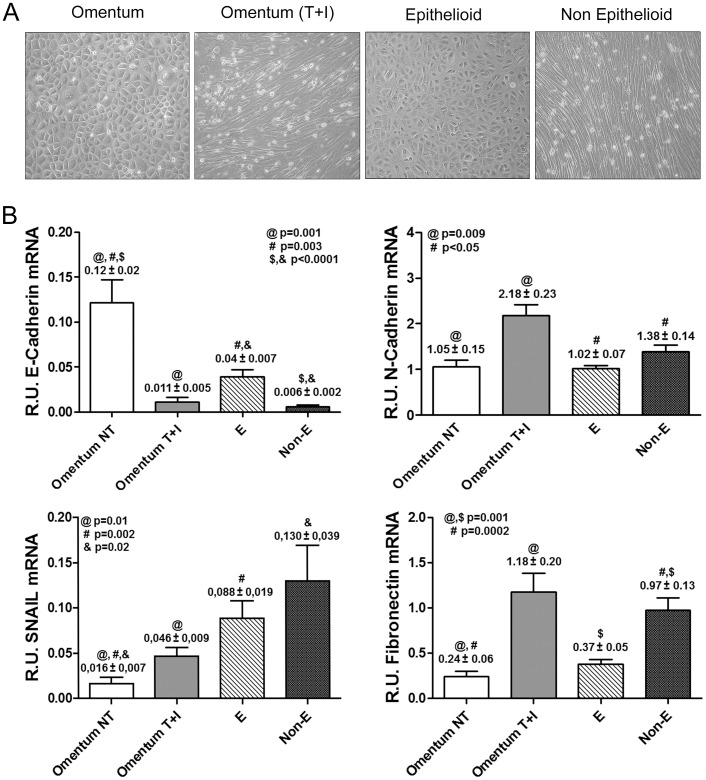
Figure 1. Characterization of MMT in vitro and ex vivo.
(A) Representative pictures of omentum-derived MCs, either untreated or treated with TGF-β1 and IL-1β (MMT in vitro), and the two morphologies observed in confluent cultures of effluent-derived MCs: epithelioid and non-epithelioid. (B) Transcript levels of mesenchymal markers were analyzed by quantitative RT-PCR (n = 11 Omentum, 11 Omentum T+I, 30 E and 21 Non-E). Results show down-regulation of E-cadherin and up-regulation of snail expression during both in vitro and ex vivo MMT. The histograms also show a significant up-regulation of N-cadherin and fibronectin expression in mesenchymal MCs compared to omentum and epithelioid MCs. Data are depicted as mean value ± SE. Symbols show statistical differences between groups.

