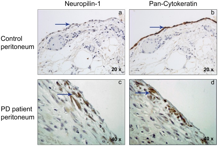
Figure 6. Nrp-1 immunohistochemical analysis in peritoneal human biopsies.
The expression of Nrp-1 and the mesothelial marker cytokeratin was analyzed in human peritoneal specimens by immunohistochemistry. Positive cells for antibodies used (Nrp-1 and Cytokeratin) show brown staining. Nuclei are counterstained in blue. (a, b) Control peritoneal tissue, with a conserved mesothelial cell monolayer showing an epithelioid morphology (with a 20X objective). These cells show weak expression of Nrp-1 and a marked staining for cytokeratin (arrows). No expression of these proteins was observed in the submesothelial area (region under mesothelial monolayer) (c, d) Fibrotic tissue sample from PD patient showing the loss of mesothelial monolayer and invading spindle-like mesothelial cells in submesothelial area (with a 40X objective). These cells present a strong staining for Nrp-1 (c), and are also positive for cytokeratin (d) (arrows). Pictures are representative of 5 cases of PD patient samples and 4 of control samples.

