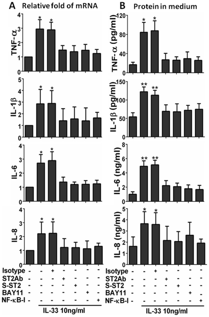
Figure 3. ST2 and NF-κB signaling pathways were involved in IL-33 induced inflammatory response.
The HCECs were exposed to IL-33 (10 ng/ml) with prior incubation in the absence or presence of isotype IgG (5 µg/ml), ST2Ab (5 µg/ml), Soluble ST2 protein (S-ST2, 10 ng/ml), BAY11-7082 (10 µM) or NF-κB activation inhibitor quinazoline (NF-κB -I, 10 µM) for 1 h. The cultures treated by IL-33 for 4 h were subjected to RT-qPCR to measure mRNA (A), the cultures treated for 48 h were used to evaluate protein in medium supernatants by ELISA (B). Results shown are the mean±SD of four independent experiments. *P<0.05; **P<0.01, n = 4.

