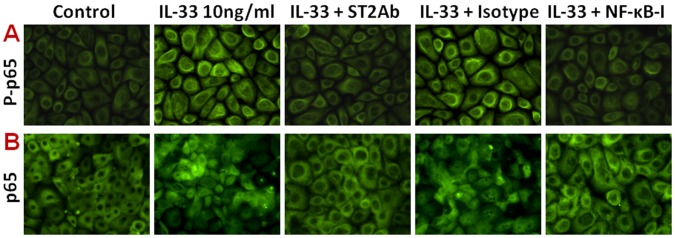
Figure 4. NF-κB activation was induced by IL-33 and inhibited by ST2 antibody and NF-κB activation inhibitor quinazoline (NF-κB-I) in HCECs.

The HCECs were exposed to IL-33 (10 ng/ml) with prior incubation in the absence or presence of ST2Ab (5 µg/ml), isotype IgG (5 µg/ml) or NF-κB-I (10 µM) for 1 h. The cells treated by IL-33 for 1 or 4 h in 8-chamber slides were used for immunofluorescent staining with rabbit antibody against phosphor-p65 (P-p65, A) or total p65 (B), respectively. The representative images were from three independent experiments. Magnifications 400X.
