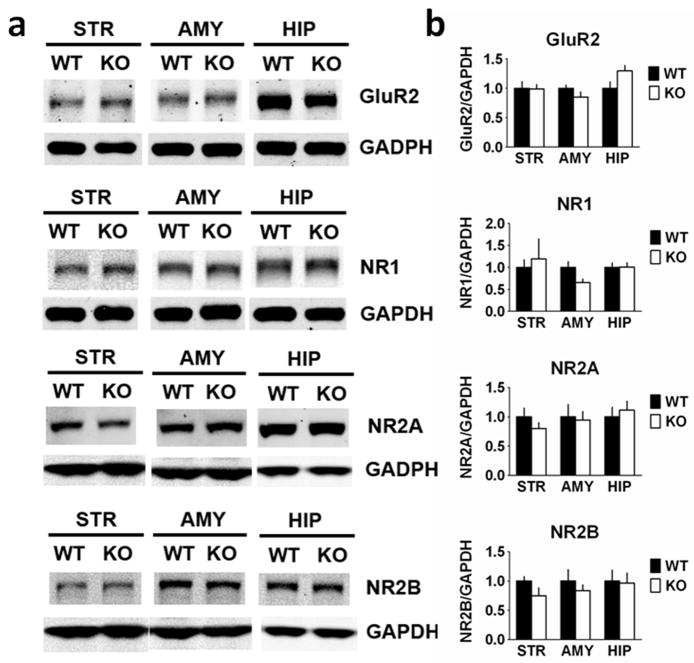
Fig. 6.
Glutamate receptor type 2 (GluR2) and NMDA receptor (NR) expression is unchanged in RCS knockout (KO) mice. Proteins from striatal (STR), amygdala (AMY) and hippocampal (HIP) extracts were analysed by SDS–PAGE and immunoblotting using GluR2, NMDA receptor type 1 (NR1), NMDA receptor type 2A (NR2A), NMDA receptor type 2B (NR2B) and glyceraldehyde 3-phosphate dehydrogenase (GAPDH) antibodies. Representative immunoblots are shown in (A), and quantitation is shown in (B) and (C). Expression levels of GluR2, NR1, NR2A and NR2B were normalized to that of GAPDH. Error bars indicate SEM, n = 5–7 per group. No differences in expression levels of any of these other glutamate receptor subunits were found in RCS KO mice (Table 1). WT, wild-type.

