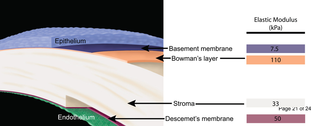
Fig. 1.
A schematic depicting the layers of the human cornea and the corresponding elastic modulus values obtained from atomic force microscopy: the epithelium, the anterior basement membrane (7.5 kPa), Bowman’s layer (110 kPa), the stroma (33 kPa), Descemet’s membrane (50 kPa) and the endothelium. Illustration (without elastic modulus values) reprinted with permission from (Last et al., 2009).

