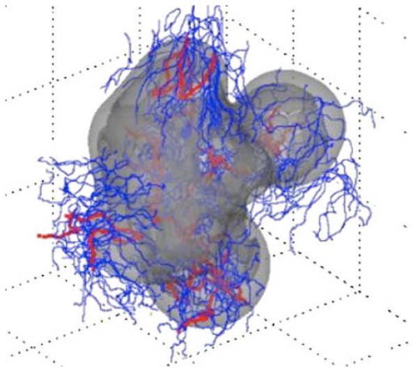
Fig. 3. 3D computer model predicts gross morphologic features of a growing glioblastoma.
Viable (light gray) and necrotic (dark gray) tissue regions and vasculature (mature blood-conducting vessels in red; new non-conducting vessels in blue) are shown. The simulations reveal that the morphology is affected by neovascularization, vasculature maturation, and vessel cooption. Adapted with permission from (67).

