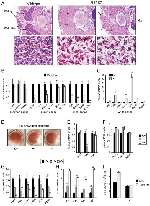
Figure 7. Ebf2 is required for BAT development in mice.
(A) Hematoxylin and Eosin (H&E) staining of representative sections from the interscapular region of wildtype and Ebf2-deficient embryos at E18 dpc. (B, C) BAT from Ebf2+/− and Ebf2−/− embryos (E18) was examined by real-time PCR analysis for mRNA levels of: general adipocyte genes, brown-specific genes and mitochondrial genes (B); and WAT-selective genes (C) (n=5–8 embryos/genotype). (D) Brown fat precursors from Ebf2+/+, Ebf2+/− and Ebf2−/− embryos were differentiated into adipocytes in culture and stained with oil-red-o. (E–H) Above cultures were analyzed by real-time qPCR for their expression levels of: Ebfs (E); general adipocyte markers (F); brown-specific genes (G); and white-selective genes (H) (n=3 pools [> 5 mice per pool]). (I) Oxygen consumption in cultured brown adipocytes from Ebf2+/− and Ebf2−/− before and after stimulation with norepinephrine (NE) (mean ± SD; n=3). Expression data are: mean ± SD, * p <0.05; ** p <0.01.

