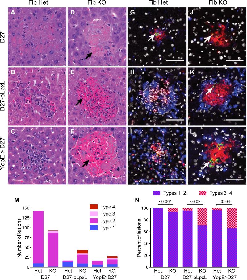
Figure 5. Fibrinogen reduces hemorrhagic pathology and increases neutrophil viability during immune defense against Y. pestis.
Fibrinogen-heterozygous mice (Fib Het) and fibrinogen-deficient mice (Fib KO) were challenged with (A,C,D,F,G,I,J,L) 2×10⁵ CFU Y. pestis strain KIM D27 or (B,E,H,K) 2×10⁶ CFU Y. pestis strain D27-pLpxL and hepatic tissue was collected four days after initiating infections. Where indicated (C,F,I,L), mice were immunized with YopE69-77 prior to challenge. (A-F) Representative paraformaldehyde-fixed samples stained with hematoxylin and eosin stained sections (400x). Hemorrhagic pathology (collections of red blood cells; black arrows) was evident in Fib KO mice challenged with D27-pLpxL (E) and in the YopE-immunized Fib KO mice challenged with KIM D27 (F). (G-L) Representative fresh-frozen samples stained with anti-F1 to identify Y. pestis (green; white arrows), F4/80 to identify macrophages (blue), anti-Ly6G to identify neutrophils (red), and Hoescht dye to identify nuclei (white). The white bar depicts 50 um. (M,N) Scoring of lesion types for mice (n=5/group) described in A-F. The Material and Methods section describes the criteria used for assigning lesion types. Typical type 1 lesions are shown in B and C; a typical lesions of type 2 is shown in A; a typical type 3 lesion is shown in D; and typical type 4 lesions are shown in E and F. The graphs depict (M) the number of lesions and (N) the percentage of hemorrhagic lesions (type 3 and 4). Statistical significance was analyzed using Student's t-tests.

