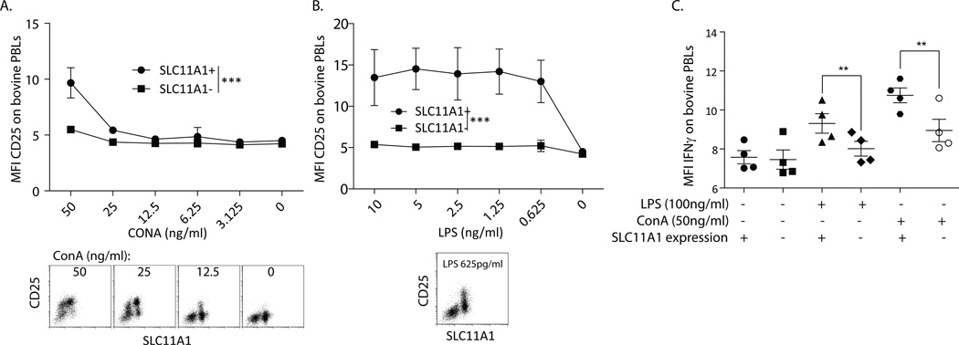
Figure 3.
SLC11A1 was strongly associated with activation of bovine cells. Bovine PBMCs were activated for 24 hours with diminishing concentrations of ConA or LPS and stained for SLC11A1 and CD25 or IFN-γ. A. SLC11A1+ cells were slightly more prone to activation (CD25 expression) by low doses of ConA, significance as calculated by 2-way ANOVA. Representative FACS plots are shown below from high to low ConA concentrations. B. Upon stimulation with very low doses of LPS, only SLC11A1+ cells were induced to express CD25. SLC11A1+ and SLC11A1− cells were significantly different as calculated by 2-way ANOVA. A representative FACS plot is shown. C. Bovine SLC11A1+ cells expressed significantly more intracellular IFN-γ. Bovine PBMCs were stimulated with 100 ng/ml LPS or 50 ng/ml ConA for 12 hours, treated with Brefeldin A for 6 hours, and then fixed and stained for SLC11A1 and IFN-γ expression. Only SLC11A1+ cells expressed IFN-γ in response to LPS stimulation and were significantly more responsive to ConA (Students t test). These data represent at least 3 repeat experiments performed with at least 3 individual calves per experiment. Error bars represent standard error, ***p<0.001, **p<0.01.

