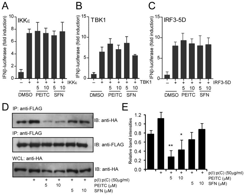
Fig. 4. PEITC interferes with TLR3 oligomerization.
HEK293 cells in 96-well plates were cotransfected with IFNβ firefly luciferase and β-actin Renilla luciferase, together with 20 ng IKKε (A), 20 ng TBK1 (B) and 40 ng IRF3-5D mutant (C) for 24 h. The transfected cells were treated with PEITC and SFN at the indicated concentrations for an additional 8 h. The results were expressed as fold induction of IFNβ luciferase activity for IKKε, TBK1 or IRF3-5D transfected cells relative to those of vector transfected controls after normalization to Renilla luciferase. The results were representative of two similar experiments. No significant difference between PEITC or SFN treated samples and non-treated samples was found by one-way ANOVA analysis. (D) HEK293 cells stably expressing FLAG-TLR3 and HA-TLR3 were treated with PEITC and SFN at the indicated concentrations for 1h and then stimulated with 50 μg/mL p(I):p(C) for another 3 h. Cell lysates were immunoprecipitated with anti-FLAG and the immunoprecipitates together with cell lysate control detected by immunoblotting for HA-TLR3. (E) Band intensities of each lane from the top panel in (D) were quantified and normalized with corresponding those of the bottom panel (anti-HA bands in whole cell lysates) and plotted as relative intensities. The plotted graph represents the average band intensities plus standard errors from two similar experiments. * p < 0.05 and ** p < 0.01, the ITC treated samples versus p(I):p(C) control sample performed by one-way ANOVA analysis.

