
Figure 2. Anti-IL-4 treatment of CD8-deficient recipients of CD8+ T cells prevents restoration of AHR and inflammation.
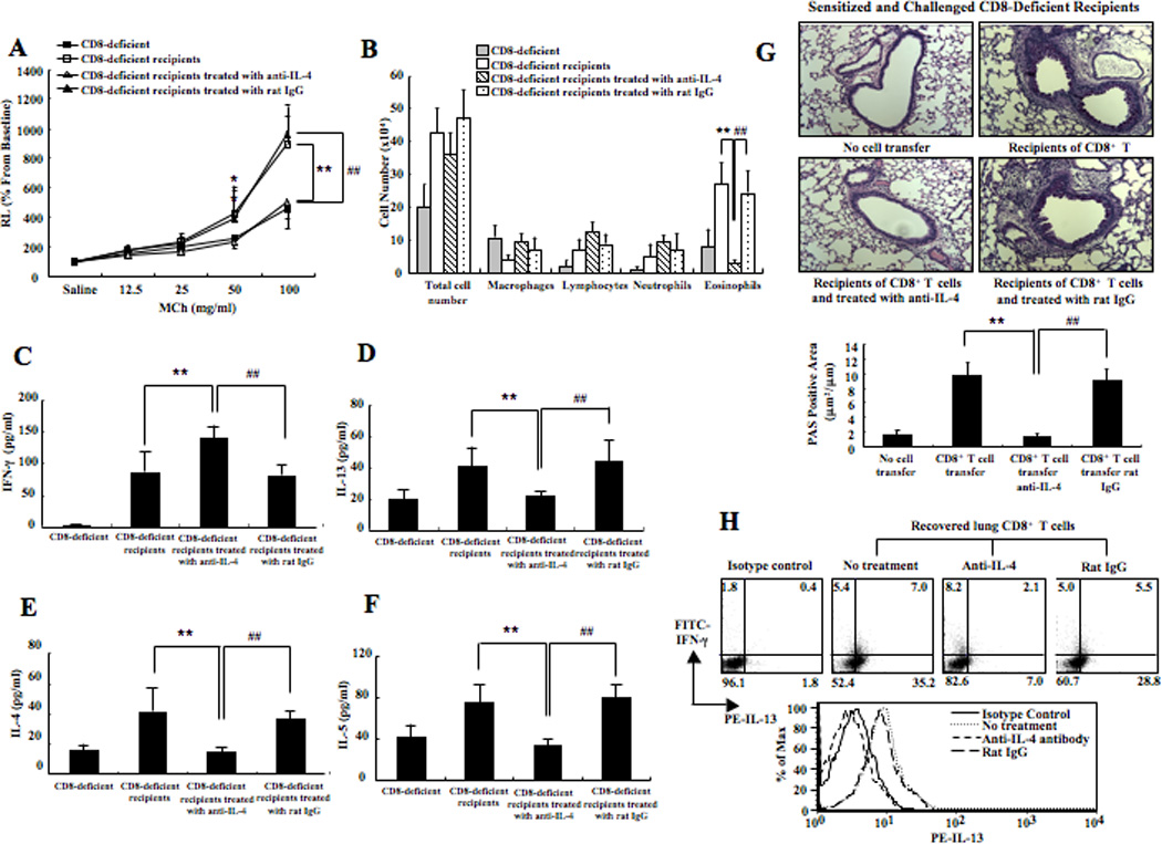
(A) Changes in airway resistance (RL) were measured in response to increasing concentrations of methacholine. (B) Cell composition in BAL fluid. Cytokine levels in BAL fluid. The top 2 graphs show IFN-γ (C) and IL-13 (D) levels and the bottom 2 graphs show IL-4 (E) and IL-5 (F) levels. (G) Representative photomicrographs of lung histology (×200). Quantitative analysis of goblet cells was as described in Materials and Methods. (H) IFN-γ and IL-13 expression in recovered lung CD8+ T cells. Data (mean±SEM) were from at least 6–10 mice. **p<0.01 and *p<0.05 compared to sensitized and challenged CD8-deficient recipients of 5×10⁶ IL-2-differentiated CD8+ T cells group. ##p<0.01 compared to sensitized and challenged CD8-deficient recipients of 5×10⁶ IL-2-differentiated CD8+ T cells treated with an isotype control.