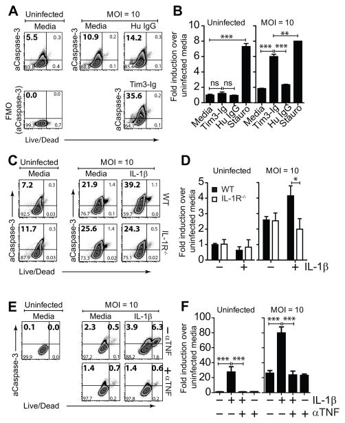
Figure 5. Tim3 and IL-1β induce caspase-3 activation.
(A) Mtb infected and uninfected thioglycolate-elicited Mϕ were cultured either alone or in the presence of 10 μg/mL Tim3-Ig or Hu IgG (Control). 24 h later, intracellular expression of active caspase-3 (aCaspase-3) in CD11b+ F4/80+ Mϕ was assessed by flow cytometry. (B) Fold induction of active caspase-3 compared to untreated uninfected Mϕ is graphically plotted. Stauro, staurosporine [1 μM], positive control. (C) Mtb infected and uninfected thioglycolate-elicited Mϕ were cultured either alone or in the presence of 10 ng/mL IL-1β. 24 h later, intracellular expression of active caspase-3 in CD11b+ F4/80+ Mϕ was assessed by flow cytometry. (D) Fold induction of active caspase-3 over untreated uninfected Mϕ is graphically plotted for uninfected and Mtb infected WT and IL-1R−/− Mϕ. (E) Mtb infected and uninfected human MDM were cultured either alone or in the presence of 10 ng/mL IL-1β with and without 25 μg/mL anti-TNF neutralizing antibody (αTNF). 24 h later, intracellular expression of active caspase-3 in CD14+ Mϕ was assessed by flow cytometry. (F) Fold induction of active caspase-3 compared to untreated uninfected Mϕ is graphically plotted. Data is representative of 2 (A, B, E, F) and 3 (C) independent experiments. Pooled data from 3 independent experiments is shown in (D). *p<0.05, **p<0.01, ***p<0.001, one-way anova compared to conditions: (B) Tim3-Ig and Staurosporine, (D) IL-1β treated WT Mϕ or (F) IL-1β treated human MDM. Bars indicate mean ± SEM from 3 replicate cultures.

