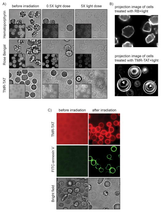
Figure 2.
TMR-TAT-mediated photohemolysis is accompanied by cell shrinkage but photolysis mediated by RB or hematoporphyrin (HP) is not. A) Bright field and fluorescence imaging of RBCs treated with HP, RB, or TMR-TAT. Images were acquired at 0.5x light dose (light dose that yields 50% photohemolysis) and 5x light dose (a light dose 10 times that required for 50% photohemolysis). Insert: fluorescence images. B) Projection images of cells treated with RB or TMR-TAT and light. The fluorescence images of the cells exposed at 0.5x, 2x and 5x light dose were super-imposed in a single overlay image. The fluorescence signal corresponds to either RB or TMR-TAT binding to the surface of lysed cells. In the case of cells treated with RB, the projection image is identical to the 0.5x image, indicating that the morphorlogy of the cells is unchanged during light exposure. In contrast, the projection image of the cells treated with TMR-TAT shows concentric circles that correspond to the cell membrane shrinking with increasing light exposure (white arrows indicate the direction of membrane shrinking with increasing irradiation). C) AnnexinV staining of membrane of RBCs lysed with TMR-TAT upon irradition. FITC-annexinV was added before or after lysis of RBCs with TMR-TAT and light irradiation.

